Membership NO : 1 Posts : 1672 Join date : 2011-03-27
Subject: Atopic Dermatitis in Emergency Medicine Mon Mar 28, 2011 9:58 am
F [center]Atopic Dermatitis in Emergency Medicine
Introduction Background Eczema, or atopic dermatitis, is a common inflammatory disease of the skin. The condition often has its start in childhood and follows a variable and sometimes unremitting course. Historically, this disease has been considered a part of a triad of "atopy" that included asthma and allergic rhinitis, though this association has recently come into question. Although not a cause of significant mortality, the visible and chronic nature of eczema can be a source of emotional stress. Pathophysiology
The precise mechanism for the development of eczema is unknown. Whether the clinical manifestations of atopic dermatitis (AD) are the result of violation of the epidermis and the subsequent contact between environmental irritants and immune cells, or the reverse sequence, is debatable. Nonetheless, the epidermis is the first line of defense between the body and the environment and, when intact, shields the body from a variety of irritants, allergens, and microbes. This barrier, which is maintained by differentiated keratinocytes and structural proteins, can be compromised by inheritance, trauma, decreased humidity,change in pH, and infection. Atopic skin additionally has diminished ability to maintain water; this dry skin leads to scratching, which further contributes to the release of proinflammatory mediators. Eczema is a biphasic T-cell – mediated disease: TH2 is more prevalent in the acute phase, and TH1 predominates in the chronically affected skin.1 Patients with atopic dermatitis have elevated serum IgE levels, peripheral eosinophilia, and overall greater numbers of immune mediators and cytokines. Frequency Atopic dermatitis is the most common inflammatory skin disease in children, affecting up to 17% of the pediatric population in the United States, with increasing prevalence over the past several decades. United States Prevalence of atopic dermatitis ranges from approximately 7-17% in children.2 A small percentage of affected children will have the disease into adulthood. International Studies in Japan and Northern Europe have found similar prevalence, with industrialized and westernized nations noting increasing trends of patients with atopic dermatitis. Mortality/Morbidity Mortality is not associated with atopic dermatitis. The impact of eczema is hard to measure but has real personal, social, and financial consequences. The burden includes but is not limited to professional fees, hospitalization, pain/suffering, social isolation, poor self-esteem, and work and/or school performance or absence. Additionally, patients suffering from atopic dermatitis are prone to bacterial superinfection. Sex Data shows a slightly increased prevalence of atopic dermatitis in female children.
Age Atopic dermatitis predominantly affects infants and young children. Eczema is apparent in the first year of life in 60% of cases, and its onset is before 5 years in 75% of cases.3 Onset of eczematous appearing disease in adulthood should lead the physician to consider another diagnosis. A triphasic course of atopic dermatitis across the lifespan has been proposed. Phase I develops before IgE sensitization has taken place and occurs mostly in infants who are likely genetically predisposed to the disease. Phase II involves IgE sensitization to food, environmental antigens, or both. Phase III is the product of chronic scratching and is characterized by the formation of IgE autoantibodies against proteins of keratinocytes and endothelial cells.
Clinical History The hallmarks of atopic dermatitis are intense pruritus, chronic eczematous skin lesions, and epidermal thickening and hypertrophy.
The emergency physician often is the first to diagnose atopic dermatitis. The most common presentation is that of infants, usually younger than 6 months, brought in by their parents for a persistent rash. Before coming to the ED, the parents may have tried a number of over-the-counter and home remedies. Parents usually report that the rash has waxed and waned for months with a history of dry skin since birth.
Clinicians should inquire about a family history of asthma, hay fever, allergy, and other atopic diseases. Patients with pertinent medical or family history of such disease tend to have a worse prognosis.
Parents may also give a history of poor sleep or increased irritability in the patients, which is due to the desire to scratch the skin during sleep. Atopic dermatitis begins with intense pruritus, leading the patient to scratch, which results in the characteristic rash.
Atopic dermatitis typically is not associated with fever or other constitutional symptoms, and the presence of these should prompt the clinician to look for bacterial superinfection.
Physical Atopic dermatitis is a spectrum of disease that varies in presentation, severity, and distribution. Eczema defies a simple definition as the disease has differing characteristics depending on the age of the patient and the stage of the disease course.
Lesions may be acute, subacute, or chronic, each with a characteristic appearance. Lesions from one stage can convert into another stage at any time due to processes such as manipulation, irritation, allergy, or infection.
Acute lesions are intensely itchy and present as vesicles and blisters with intense redness.
Subacute disease is characterized by slight-to-moderate itching, pain, stinging, burning and redness, scaling, and fissuring of the skin with a parched and scalded appearance.
Chronic eczematous inflammation demonstrates thickened skin, accentuated skin lines, excoriations, and fissuring accompanying a moderate-to-intense itch.
The pattern of skin manifestations also differs across the lifespan.
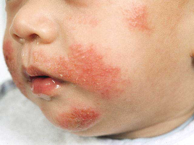
In infantile atopic dermatitis, pruritic, red, scaly, and crusted lesions are typically found on the extensor surfaces and cheeks or scalp, with severe cases possibly presenting with vesicles, serous exudates, or crusting. The diaper area is protected and usually spared.
</li>
Irritation around mouth of an infant with atopic dermatitis.
[/url]Irritation around mouth of an infant with atopic dermatitis.
The lesions in the childhood stage have less exudation; the skin often demonstrates lichenified plaques in a flexural distribution, commonly antecubital and popliteal fossae, volar aspect of the wrists, ankles, and neck.
</li>
Flexural involvement in childhood atopic dermatitis.
Flexural involvement in childhood atopic dermatitis
</li>
Product Description This newly reorganized and expanded Second Edition of the revolutionary reference guide, Atopic Dermatitis, is the only available text devoted to discussing all aspects of this common disease. Appealing to dermatologists, allergists, family practitioners, and pediatricians in daily practice, as well as physician scientists, medical students, and experimental investigators, this stand-alone guide addresses recent breakthrough discoveries in genetics and new milestones in immunological research in dermatology. New highlights in the Second Edition:
12 brand new chapters that encompass the latest research
the latest advances, including regulatory and Th17 cells in atopic dermatitis, cytokines and chemokines, virus infections, autoantigens, specific immunotherapy, the immune system and atopic dermatitis, and neuromediators and the pathophysiology of pruritus
coverage of the most recent areas of advancement: pathophysiology, mechanisms and cellular aspects, epidemiologic, genetic, and immunologic aspects of atopic dermatitis
=======================================
GET IT HERE
================================
====================== Related Topics: A Practical Manual Handbook of Dermatology Free Download Cosmetic Dermatology: Principles and Practice Free Download Radiation Therapy for Skin Cancer Free Download Color Atlas of Chemical Peels Free Download The Manual of Dermatology Free Download Dermatology in Clinical Practice Free Download Hair and Scalp Diseases Free Download
john
Membership NO : 1 Posts : 1672 Join date : 2011-03-27
Subject: Re: Atopic Dermatitis in Emergency Medicine Mon Mar 28, 2011 9:59 am
Adult eczema has a similar distribution to that in childhood atopic dermatitis but is increasingly localized and lichenified with thickened skin, increased skin markings, and excoriated and fibrotic papules.
Certain characteristic patterns are worth mentioning.
Eczema that appears as one or several coin-shaped plaques is called nummular eczema.
Plaques with prominent skin lines are referred to as lichen simplex chronicus. These lesions are characterized by intense pruritus that ceases when pain replaces itch.
Diagnosis of atopic dermatitis is made by observing representative clinical features of the disease. The Hanifin and Rajka diagnostic criteria, which consist of 4 major and 23 minor criteria has traditionally been used, but it is time consuming and not manageable. The UK working group on atopic dermatitis has the following criteria for diagnosis, which has been most extensively validated in clinical trials.4 Evidence of itchy skin with 3 more of the following:
History of skin crease involvement
Presence of generally dry skin within in the past year
Symptoms in a child beginning before the age of 2 years
Visible involvement of dermatitis involving flexural surfaces
The complete Hanifin and Rajka criteria are included below:
Major criteria (need 3 or more)
Pruritus
Typical morphology and distribution
Flexural lichenification in adults
Facial and extensor involvement in infants and children
Dermatitis - Chronically or chronically relapsing
Personal or family history of atopy (asthma, allergic rhinitis, atopic dermatitis)
Minor criteria (need 3 or more)
Cataracts
Cheilitis
Conjunctivitis - Recurrent
Eczema - Perifollicular accentuation
Facial pallor or erythema
Food intolerance
Hand dermatitis - Nonallergic
Ichthyosis
IgE - Elevated
Immediate (type I) skin test reactivity
Infections (cutaneous)
Dennie-Morgan infraorbital fold
Itching when sweating
Keratoconus
Keratosis pilaris
Nipple dermatitis
Orbital darkening
Palmar hyperlinearity
Pityriasis alba
White dermographism
Wool intolerance
Xerosis
Causes
Atopic dermatitis is a complex genetic disease that results from an array of gene-gene and gene-environment interactions. Most experts believe that atopic dermatitis has a genetic basis. This is supported by twin studies and chromosome studies that suggest the trait might be inherited via a maternal gene located on chromosome 11. Clinical studies have also shown a higher risk of atopy in children with maternal atopy than in children with paternal atopy.5
Two theories have been proposed to explain the manifestations of atopic dermatitis. Atopic dermatitis was traditionally thought to be caused by an innate immunologic disturbance leading to IgE sensitization, which later results in disruption of the epithelial barrier, though this supposed mechanism is falling out of favor. Alternatively, it is thought that skin breakdown precedes the inflammatory process and an intrinsic epithelial cell defect leads to barrier disruption of the skin and that immunologic imbalance is an "epiphenomenon".1 Genetic defects in filaggrin, a group of structural proteins, have been cited as a major cause of atopic dermatitis.6,7 The upregulation of a protease stratum corneum chymotryptic enzyme is also being investigated in the cause of atopic dermatitis.
Chronic eczema is a disease that is somewhat behaviorally mediated. Skin thickening and plaque formation is dependent on habitual scratching. ==============================
GET IT HERE
====================== Related Topics: A Practical Manual Handbook of Dermatology Free Download Cosmetic Dermatology: Principles and Practice Free Download Radiation Therapy for Skin Cancer Free Download Color Atlas of Chemical Peels Free Download The Manual of Dermatology Free Download Dermatology in Clinical Practice Free Download Hair and Scalp Diseases Free Download
john
Membership NO : 1 Posts : 1672 Join date : 2011-03-27
Subject: Re: Atopic Dermatitis in Emergency Medicine Mon Mar 28, 2011 9:59 am
Workup
Laboratory Studies
Laboratory testing, including IgE levels, is not necessary or recommended in the evaluation of suspected atopic dermatitis. Procedures
Pathologic findings of biopsy samples include spongiosis with a severely damaged stratum corneum, with hyperproliferation in advanced cases.
Histological studies show an inflammatory infiltrate consisting of memory predominantly CD4 T lymphocytes. Chronic lesions also display increased mast cells, eosinophils, and IgE-bearing Langerhans cells.
Treatment
Emergency Department Care
Many patients with atopic dermatitis (AD) present to the ED during acute exacerbation. Therapy is targeted toward alleviation of pruritus and prevention of scratching. ED physicians must also look for signs and symptoms of bacterial superinfection and treat accordingly.
Practice guidelines on the management of atopic dermatitis are available from the Joint Task Force on Practice Parameters for Allergy and Immunology.8
Skin care
In the acute setting patients should be instructed to bathe once-to-twice daily using mild soaps (eg, Dove). There is no preference over showers or baths, whichever makes the patient most comfortable.
The patient should dry quickly and immediately (within 3 min) lubricate the skin. Many creams and lotions are available, and the optimal one is the greasiest the patient can tolerate.
Creams (eg, Eucerin, Cetaphil) are preferred over lotions, as they have lower or no water content and will not evaporate off of the skin during the day. Parents may use petroleum jelly on infants, but most children and adults will not tolerate the texture.
</li>
Topical steroids
Acute attacks should be treated by mid-high strength topical steroids for up to 2 weeks. Medium-to-high potency topical steroids should not be used on the face or neck area because of the potential adverse effects. These are preferred over low-mid strength medications, as they better control exacerbations. Patients should apply the ointment within 5 minutes of twice-daily bathing.
Inform the patient about adverse effects of topical steroids (eg, atrophy, hypopigmentation, striae, telangiectasia, thinning of the skin).
</li>
Antihistamines: Physicians have been prescribing antihistamines for years to control the pruritus associated with acute atopic dermatitis. Little evidence exists that antihistamines help with the itching in an awake patient; however, the use of sedating antihistamines is supported to control scratching while the patient is asleep.9
Systemic steroids: The use of systemic steroids in the treatment of acute exacerbation of atopic dermatitis is controversial. Most authors reserve oral prednisone (at least 20 mg/d for 7 d) for the most severe cases, although it seems the disease quickly relapses once the medication is discontinued. Patients also tend to discontinue topical steroid creams and other treatment as they feel better, which contributes to the relapse after oral steroids are done.
Topical calcineurin inhibitors: Topical calcineurin inhibitors (pimecrolimus 1% and tacrolimus 0.03%, 0.1%) are available for patients older than 2 years. These medications may be used all over skin surfaces (including face, neck, and hairline) because they do not have the side effects seen with topical steroids. Evidence supports the twice-daily use of these creams during acute exacerbation of atopic dermatitis, and some evidence exists to support use up to 4 years. The long-term side effects (including the possibility of increased risk for malignancy) have not fully been elucidated. For these reasons, the US Food and Drug Administration (FDA) does not recommend long-term use yet. Side effects of tacrolimus include burning and stinging on broken skin.
Oral immunosuppressive agents: Patients with refractory atopic dermatitis may benefit from oral immunosuppressive agents, such as cyclosporine A. This medication is effective in treating severe atopic dermatitis in the acute setting. It is not recommended for long-term use.
Phototherapy with PUVA, UVA, or UVB is successful in controlling atopic dermatitis but is expensive and may lead to increased risk of melanoma and nonmelanoma skin cancer. Studies evaluating the role of several proposed disease-modifying agents continue to be conducted. Results of studies on IL-4 neutralizing antibody and mast cell depleters are promising, whereas the evidence for probiotics,10,11 primrose oil, sodium cromolyn, topical caffeine, and dietary exclusion is inconclusive and requires more research.
Consultations
In cases of uncertain diagnosis or severe atopic dermatitis that is resistant to conventional therapy, patients may be referred to a specialist such as a dermatologist or an allergist
==================
GET IT HERE
john
Membership NO : 1 Posts : 1672 Join date : 2011-03-27
Subject: Re: Atopic Dermatitis in Emergency Medicine Mon Mar 28, 2011 9:59 am
Follow-up
Further Inpatient Care
Few patients with atopic dermatitis will require hospitalization. Patients with cellulitis or severe secondary infection may require intravenous antibiotics and sedation.
Further Outpatient Care
Atopic dermatitis is chronic and relapsing. The main goal of treatment is control of the disease, not a cure. Patient education about management of flare and recognition of superinfection is paramount.
Inpatient & Outpatient Medications
Few patients with atopic dermatitis will require hospitalization. Patients with cellulitis or severe secondary infection may require intravenous antibiotics and sedation.
Deterrence/Prevention
Breastfeeding has indisputable benefits in infant nutrition, but no strong evidence suggests a protective effect against the development of atopic dermatitis, even in children with a family positive history.12
The mainstay of treatment of atopic dermatitis is prevention of outbreaks. Patients should continue to take short showers/baths followed by immediate hydration of skin with emollients even during rash-free times. They should also continue to avoid any triggers that may exacerbate atopic dermatitis.
Complications
Excoriations secondary to itch predispose to infection and can be recognized by the accumulation of serum, crust, and purulent material. Development of vesicle and/or pustules in patients with known atopic dermatitis should initiate a search for bacterial or viral superinfection; appropriate antibiotics or antivirals should be started immediately. Patients with atopic dermatitis are uniquely susceptible to herpes simplex, which may occasionally progress to a Kaposi’s varicelliform eruption; physicians and patients should be particularly vigilant for this condition. This rare complication is characterized by generalized involvement, systemic toxicity, and even death. In these cases, the patient should be treated with oral acyclovir and monitored closely. Topical corticosteroids and/or occlusive dressings are best, at least temporarily, discontinued.
Exfoliative erythroderma is another rare complication, occurring in less than 1% of patients with atopic dermatitis. Exfoliative erythroderma demonstrates a marked progression caused by widespread Staphylococcus aureus or herpes simplex superinfection and can be life threatening if it is complicated by high-output cardiac failure and heat loss.
Atrophy or striae occur if fluorinated corticosteroids are used on the face or in skin folds.
Systemic absorption of steroids may occur if large areas of skin are treated, particularly if high-potency medications and occlusion are combined.
Prognosis
About 90% of patients with atopic dermatitis have spontaneous resolution by puberty.
Adults who continue to have atopic dermatitis usually have localized dermatitis (eg, chronic hand or foot dermatitis, eyelid dermatitis, lichen simplex chronicus).
Unfavorable prognostic factors for atopic dermatitis (in order of relative importance) include the following:
Persistent dry or itchy skin in adult life
Widespread dermatitis in childhood
Family history of atopic dermatitis
Associated bronchial asthma
Early age at onset
Female gender
</li>
The objective when treating a patient for atopic dermatitis is control of exacerbations, not elimination of the disease. Using the above treatment modalities, patients and their families can hope to minimize the frequency and severity of outbreaks.
Patient Education
Given the chronic nature of atopic dermatitis and patients' concerns about appearance, emotional support and psychological counseling may be helpful. Physicians need to be sensitive to the needs of patients and their families
Membership NO : 1 Posts : 1672 Join date : 2011-03-27
Subject: Re: Atopic Dermatitis in Emergency Medicine Mon Mar 28, 2011 10:00 am
Multimedia
(Enlarge Image) Media file 1: Typical atopic dermatitis on the face of an infant.[ CLOSE WINDOW ]<blockquote></blockquote>Typical atopic dermatitis on the face of an infant.
(Enlarge Image) Media file 2: Flexural involvement in childhood atopic dermatitis.[ CLOSE WINDOW ]<blockquote></blockquote>Flexural involvement in childhood atopic dermatitis.


 [/url]Irritation around mouth of an infant with atopic dermatitis.
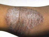
 Flexural involvement in childhood atopic dermatitis
 (Enlarge Image)
 </blockquote>Typical atopic dermatitis on the face of an infant.