Medical Book
Would you like to react to this message? Create an account in a few clicks or log in to continue.

Medical Book

Buy Textbooks | Autoclaves | stethoscopes | Buy Books Online | Buy Medical Textbooks | Textbooks | Equipment | Nutrition | USMLE | MRCP | MRCS | Dental | Sport Medicine | Cardiology | Medical Textbook | Surgery | Pregnancy | Anatomy | Radiation | Pedia |
 
HomeLatest imagesPublicationsRegisterLog in

Share
 

 Contact Lens Complications

View previous topic View next topic Go down 
AuthorMessage
john

john

Membership NO : 1
Male Posts : 1672
Join date : 2011-03-27

Contact Lens Complications Empty
PostSubject: Contact Lens Complications   Contact Lens Complications Icon_minitimeMon Mar 28, 2011 10:50 am

Contact Lens Complications

Introduction

Contact lens–related complications range from self-limiting to sight
threatening, which require rapid diagnosis and treatment to prevent
vision loss. With millions of individuals wearing contact lenses, even a
small percentage of complications can constitute a major public health
problem. Contact lens complications are as varied as they are common,
involving the lids, conjunctiva, and all layers of the cornea (ie,
epithelium, stroma, endothelium). When reviewing complications, grouping
them according to which anatomical structure is primarily affected can
be helpful.

For illustrated demonstrations of removing different types of contact lenses, see eMedicine's Contact Lens Removal article.
Lids

Giant papillary conjunctivitis is a common complication among soft contact lens wearers.1 This complication is often addressed by changing 1 of the following 3 factors:


  1. Increasing the frequency of lens disposal
  2. Decreasing the length of contact lens wear
  3. Switching to a stronger cleaning solution
Rarely, rigid contact lenses can dislocate from the cornea and
settle into the upper fornix. If undetected, the lens may erode through
the conjunctiva and enter the soft tissues of the lid, where it can
remain relatively asymptomatic. Alternatively, the tissues around the
contact lens can become irritated and inflamed, producing a sterile
abscess. The lens foreign body can incite the formulation of
granulation tissue around the lens, encapsulating it in a cystlike
structure.

A mechanical ptosis
is occasionally the result of the mass of lens, scar, and fibrous
tissue in the lid. An embedded contact lens also can produce enough
scarring and contraction of the lid tissues to produce a lid retraction.
The contact lens need not migrate into the lid tissues to produce
ptosis. A ptotic lid can result simply from severe giant papillary
conjunctivitis (GPC).

Occasionally, ptosis can be seen in contact lens wearers without any
inflammation, lens migration, or other definite cause. Hard contact
lens wearers may develop ptosis from levator aponeurosis disinsertion
from years of repeated stretching of the lid during lens removal. A
second proposed mechanism is that the repeated trauma of the lens edge
rubbing against the palpebral conjunctiva produces chronic inflammation
and edema in the soft tissues of the lid. Because all or part of the
ptosis may resolve with discontinuation of contact lens wear, it is
recommended that patients stop wearing their lenses for a period of
time prior to surgical correction of the ptosis. Tear Film

The tear film provides a smooth and transparent refractive surface,
essential moisture, and oxygen to the epithelial cells. Tears also
contain immunoglobulins, complement, and other proteins, which help
protect against infection. The health of the ocular surface is entirely
dependent upon an adequate quantity and quality of tear film, both of
which can be altered by the presence of contact lenses.
Bacteria and debris are collected in the tear film, wiped by the lid
blink, and rinsed away from the surface of the eye. The presence of a
contact lens on the eye substantially reduces the interchange of tears
across the ocular surface. Rigid lenses reduce the tear exchange
compared to no contact lens wear. Soft lenses reduce the tear exchange
to an even greater extent and the larger the diameter, the greater the
reduction.
The effect of contact lenses on the tear film can vary from one part of
the cornea to another part of the cornea. Tear film instability exists
in the interpalpebral fissure in the periphery of the cornea, the
so-called 3- and 9-o'clock areas, in wearers of rigid contact lenses. A
rigid lens that is fit poorly often produces corneal staining at these
sites. Epithelial damage in these areas is associated with instability
and abnormalities in the mucin layer of the tear film.
In addition to the mixing of tears, the content of the tears can be
altered by the presence of contact lenses. Overnight wear increases the
levels of tear proteins compared to daily wear or no wear of contact
lenses.
Conjunctiva

Conjunctival Abnormalities
Contact allergy
A contact dermatitis hypersensitivity reaction can be produced by one
of a host of chemicals, which are found in contact lens solutions. A
typical reaction consists of marked itching with varying amounts of
injection, burning, redness, tearing, mucoid discharge, and
occasionally chemosis. In addition, the lid may become edematous and
erythematous. Cold compresses and the elimination of the offending
chemical usually relieves symptoms. A short course of topical steroids
can be used in particularly severe instances.
Giant papillary conjunctivitis
Approximately 1-3% of contact lens wearers eventually develop a symptom
complex of GPC consisting of conjunctival injection, mucoid discharge,
itching, tear film debris, coated lenses, blurred vision, excess lens
movement, and blurred vision. These symptoms may remain minimal or
progress to complete lens intolerance. The tarsal conjunctiva becomes
inflamed and hypertrophied. This inflammatory hypertrophy is
morphologically similar to the papillary hypertrophy of vernal
conjunctivitis.
The etiology of GPC is multifactorial and begins with the formation of
deposits on the surface of the lens. The constant trauma of the
blinking lid rubbing on the surface of the lens exposes the deposits to
the conjunctival lymphatic system. The antigens associated with the
deposits incite an immune response in the conjunctiva. This condition
can occur whenever a foreign substance chronically rubs the tarsal
conjunctiva, such as ocular prostheses, exposed scleral buckles, nylon
sutures, and gas-permeable contact lenses but most commonly is
associated with soft contact lenses.
Typically, papillae (0.3 mm or larger) are seen surrounded by thickened
and hypervascular conjunctiva. The hyperplastic epithelium extends
down into the underlying stoma. The epithelium is infiltrated with mast
cells, and the stroma is infiltrated with basophils and eosinophils.
The symptoms of GPC are exacerbated by anything that increases the
contact of the lens deposits with the tarsal conjunctiva, such as
increased numbers of deposits, increased size of the contact lens, and
increased wearing time, especially overnight wear.
Treatment of GPC consists of reducing the amount of contact between the
deposits and the conjunctiva. Frequent enzymatic cleaning of the
contact lenses, frequent replacement of contact lenses (disposable
lenses), reduction in wearing time, and the use of lenses that resist
deposit formation are effective treatments.
Medications that suppress the immune response also can be used. Topical
steroids also reduce symptoms; however, the risk of complications
limits their use. Topical mast cell stabilizers, such as 4% cromolyn,
have some effectiveness in reducing the symptoms of GPC. Medical
treatments generally are used for a short duration in acute
exacerbations. The most effective treatment usually is reduced wearing
time and switching to disposable contact lenses.
Contact lens–induced superior limbic keratoconjunctivitis
(CL-SLK) is an immunologic reaction in the peripheral conjunctiva
produced by contact lens wear that is similar to that seen in Theodore
superior limbic keratoconjunctivitis (SLK). It is characterized by
conjunctival thickening, erythema, and a variable amount of fluorescein
staining of the superior bulbar conjunctiva.
The keratinized epithelium loses many of its goblet cells and is
invaded by neutrophils. Foreign body sensation, photophobia, tearing,
burning, occasional itching, and reduced visual acuity due to punctate
epitheliopathy are typical symptoms of CL-SLK.
Although similar in name, CL-SLK is a separate and distinct entity from
Theodore SLK. CL-SLK can be differentiated by a lack of filaments,
minimal tarsal papillary reaction, impaired vision, and lack of
association with thyroid disease. It also is not limited to the
superior conjunctiva but can be circumferential.
CL-SLK may be caused by excessive lens movement or sensitivity to
thimerosal. Treatment consists of discontinuing contact lens wear until
the epithelium returns to normal and the symptoms resolve. Refitting
with better fitting lenses, using preservative-free solutions with a
hydrogen peroxide disinfecting system, or switching to rigid
gas-permeable (RGP) contact lenses may permit a resumption of contact
lens wear
=================================
Automatic Contact Lens Cleaner System
Contact Lens Complications 31ls1mZn1cL._SL500_AA300_Contact Lens Complications 31YGaXDe6bL._AA300_Contact Lens Complications 41dGGuEjtnL._AA300_Contact Lens Complications 41DS1G8YoYL._AA300_
READ MORE...CLICK HERE
GET IT HERE

Contact Lens Complications Buyamzon-button
save 30% order now
===================


Related Subject
ABC of Eyes (ABC Series)
Becker-Shaffer's Diagnosis and Therapy of the Glaucomas
Borish's Clinical Refraction (Benjamin, Borish's Clinical Refraction)
Ophthalmology Atlas Test Yourself Atlas in Ophthalmology
Pearls of Glaucoma Management - Feb 2010 Edition

Share
Back to top Go down
john

john

Membership NO : 1
Male Posts : 1672
Join date : 2011-03-27

Contact Lens Complications Empty
PostSubject: Re: Contact Lens Complications   Contact Lens Complications Icon_minitimeMon Mar 28, 2011 10:50 am

Corneal Epithelium

Mechanical epithelial defects
The contact lens is a foreign body that rubs across and is pressed
against the corneal epithelium with each blink, thousands of times each
day. Surprisingly, this only occasionally results in an abrasion.
Corneal abrasions from contact lens wear need to be recognized and
treated because they indicate chronic epithelial stress due to the
contact lens. Epithelial defects can allow bacteria to penetrate the
cornea, resulting in a stromal infection. Chronic corneal epithelial
trauma can stimulate subepithelial fibrosis in the absence of an
infection. The specific abrasion pattern often provides the necessary
clues to indicate what problem can be corrected to improve the comfort
and safety of the patient.
Manipulation of a contact lens during insertion and removal can
traumatize the epithelium creating painful abrasions of various shapes
and sizes. These abrasions usually heal quite rapidly with simple
lubrication or patching. Debris trapped under a contact lens or a chip
or tear in the edge of a contact lens can produce dramatic curvilinear
abrasions. Removal of the debris or replacement of the damaged
contact lens is all that is needed to treat this problem.
Punctate epithelial erosions occur commonly with contact lens wear and
have several causes. Three staining patterns are characteristic for
rigid lenses, as follows: central, peripheral, and 3- and 9-o'clock
positions. If a lens is too flat for the particular cornea, it may
produce central punctate staining. A steep cornea, such as in
keratoconus where the lens rubs on the tip of the cone, is a typical
example. A lens that is too steep for the cornea can produce peripheral
punctate staining patterns, often in a superior arcuate shape. A
poorly moving lens or one with a large optical zone may produce
superior arcuate staining.
The most common staining pattern occurs between the lens and the limbus
in the interpalpebral fissure (at the 3- and 9-o'clock positions).
This epitheliopathy is caused by the contact lens lifting the lid away
from the cornea and poor tear stability with subsequent drying of the
cornea. This often is exacerbated by an incomplete blink. A small
amount of staining (at the 3- and 9-o'clock positions) is benign, but
persistent epithelial erosions can lead to dellen formation,
neovascularization, Salzmann-type elevated lesions, and pseudopterygium
formation. This type of punctate staining is alleviated by decreasing
the distance from the lens to the limbus with a larger lens, reducing
edge lift with a thinner-edged lens or steeper fit, or refitting with
a lens that rests under the upper lid (alignment fit).
Punctate staining by soft lenses is not as common as with rigid lenses
but can occur. Soft lenses that cause excessive desiccation can cause
an inferior central or inferior arcuate pattern. Usually, these
patients have minor symptoms of mild irritation or slightly decreased
vision. Refitting with a higher water content lens or RGP lens usually
eliminates the problem.
Epithelial splitting is a common finding in asymptomatic soft contact
lens wearers. This finding often is overlooked on a routine examination
because it usually does not cause severe symptoms and may be covered
by the upper lid. Epithelial splits are horizontal, linear, white,
faintly staining epithelial defects in the superior cornea, which often
are asymptomatic during lens wear and produce mild foreign body
sensation after the lens has been removed. The splits usually heal
after the lenses have been out for 24 hours and refitting with RGP
lenses prevents recurrence.
Chemical epithelial defects
Various contact lens chemical solutions can produce a range of
epithelial defects from marked erosions to less extensive punctate
defects. Surfactant cleaning solutions that are left on the lens after
cleaning usually cause immediate pain, redness, photophobia, and
tearing upon lens insertion. These symptoms typically disappear after
1-2 days.
If hydrogen peroxide is placed on the eye, it can cause intraepithelial
and subepithelial gas bubbles. These bubbles have a dramatic
appearance and can cause significant but usually temporary vision loss.
The bubbles typically resolve without permanent sequelae within
minutes to hours. However, hydrogen peroxide can cause a permanent
refractive change by altering the shape of the cornea.
Enzyme cleaner and chemical disinfection solutions can cause more
subtle and intermittent punctate epithelial defects. This condition may
require careful investigation and systematic elimination of various
lens care products to identify and remove the offending agent. Use of
preservative-free solutions and proper use of hydrogen peroxide
disinfection usually solves this problem.

=================================
Innoxa French Blue Eye Drops Gouttes Bleues
Contact Lens Complications 31cM-8Fd5GL._SL500_AA300_
Order now and save 42%

  • Rich in natural plant extracts provides a soothing & stimulating effect
  • Helps gives a sparkle to eyes while allowing to relax & rest the eyes (good for contact wearers too)
  • The azur blue color revives the white of the eyes and gives an irresistible sparkle
  • Enhances any color eyes to a vibrant color
  • Professional photographer use on top models, also celebrities use consistently
GET IT HERE

Contact Lens Complications Buyamzon-button
save 42% now
========================================

Related Subject
ABC of Eyes (ABC Series)
Becker-Shaffer's Diagnosis and Therapy of the Glaucomas
Borish's Clinical Refraction (Benjamin, Borish's Clinical Refraction)
Ophthalmology Atlas Test Yourself Atlas in Ophthalmology
Pearls of Glaucoma Management - Feb 2010 Edition

Share
Back to top Go down
john

john

Membership NO : 1
Male Posts : 1672
Join date : 2011-03-27

Contact Lens Complications Empty
PostSubject: Re: Contact Lens Complications   Contact Lens Complications Icon_minitimeMon Mar 28, 2011 10:51 am

Hypoxia
Because the oxygen requirements of the cornea are met by direct
diffusion of oxygen from the corneal surface, the barrier of the
contact lens reduces the amount of available oxygen. Contact lens wear
(especially with a closed lid during sleep) can cause acute hypoxia.
If mild, hypoxia produces epithelial edema and temporary blurred
vision; if severe, it can cause epithelial cell death and
desquamation. Patients usually experience discomfort and remove the
contact lenses before the acute hypoxia becomes severe. Typically, the
conjunctiva is hyperemic, and the epithelium has fine punctate
defects, producing temporary decreased vision and photophobia.
Chronic hypoxia produces a variety of more subtle effects, such as
epithelial microcysts. Contact lens users who sleep in their lenses are
prone to developing epithelial microcysts. These transparent
epithelial inclusions of degenerated epithelium are about 10-15 µm,
begin in the deep epithelium, and slowly migrate anteriorly. Upon
reaching the surface, they rupture, creating depressions that pool with
fluorescein. Epithelial microcysts seldom produce any significant
symptoms other than a mild decrease in vision. Surprisingly, it takes
several weeks for the microcysts to disappear after discontinuation of
the contact lenses. Either the mitotic rate is reduced below normal
or the microcysts continue to be produced long after the contact
lenses are removed.
One of the hallmarks of chronic corneal hypoxia is superficial neovascularization, especially along the superior limbus. Neovascularization
of less than 2 mm from the limbus is not visually significant and
generally is well tolerated but is a sign of hypoxia and may be a
harbinger of more significant problems. Rarely, deep stromal
neovascularization can occur. Changing to lenses that are thinner or
contain materials with greater oxygen permeability, have greater lens
movement, and decreasing wear time (especially eliminating overnight
wear) can greatly reduce the risk of progression.
Chronic hypoxia has been implicated as a cause of the decreased
corneal sensitivity that occurs with prolonged contact lens wear and
may be partly the reason why some patients have increased comfort with
long-term wear and why they often have decreased comfort with a
change from polymethyl methacrylate (PMMA) to gas-permeable contact
lenses.
The corneal epithelium is thinner in contact lens wearers. This change
may be due to chronic hypoxia and decreased mitotic activity. In
addition to thinning of the epithelium, extended wear is associated
with decreased epithelial shedding, increased cell size, and increased
binding of Pseudomonas aeruginosa to the cell surface. All of
these effects could reduce the resistance of the cornea to bacterial
infection. The thinner epithelium poses less of a barrier to
bacterial penetration. The reduced shedding of epithelial cells
allows the attached bacteria to remain on the eye for longer periods
of time. The increased binding of bacteria, such as P aeruginosa, enables greater numbers of bacteria to attach to the epithelial surface.
The physiology of the corneal epithelium also is altered by contact
lens wear. The barrier function of the epithelium is reduced, and the
permeability to fluorescein is doubled after as little as 2 weeks of
soft contact lens wear. Similarly, rigid contact lenses can alter the
epithelial permeability.
Superficial immunologic reactions
A variety of chemicals in contact lens solutions can elicit
superficial toxic or immune reactions. The typical response is a fine
punctate keratopathy, conjunctival injection, tearing, itching, and occasionally chemosis.
The preservative, thimerosal, which is now rarely used, produced a
keratoconjunctivitis in as many as 10% of contact lens wearers who used
thimerosal-preserved products. Essentially, it has disappeared from
use, but other chemicals used as preservatives or disinfectives can
produce similar pathology, so recognition of this condition is helpful.
The earliest symptoms are mild and nonspecific (eg, foreign body
sensation, conjunctival hyperemia, variable mixed follicular-papillary
hypertrophy), which present gradually after weeks or years of
uneventful contact lens wear. The superior limbus becomes progressively
more hyperemic and a triangle of punctate keratopathy extends
downward from the involved limbus toward the central cornea. If
allowed to proceed, the epitheliopathy may progress to an opaque
pannus with microcysts.
A problem associated with the use of chemical disinfection systems and
seen with increasing frequency is the development of small, gray,
epithelial, granular opacities that resemble the epithelial opacities
of Thygeson superficial punctate keratopathy.
The round, gray-white granules appear to be on the surface of the
epithelium and are scattered randomly across the cornea. They are
similar to Thygeson superficial punctate keratopathy, but they tend to
be small and stain less intensely with fluorescein. These opacities
are associated with symptoms of foreign body sensation, tearing,
photophobia, lens intolerance, and conjunctival injection. The symptoms
resolve over a few days after the chemical disinfecting solution is
discontinued.
Thimerosal and other chemicals used in disinfection systems also can
produce subepithelial infiltrates similar to those seen in adenoviral
conjunctivitis. Changing to a preservative-free hydrogen peroxide
based disinfection system or to gas-permeable lenses allows these
deposits and infiltrates to resolve. However, it may take weeks for
the pathology to disappear.
Corneal Stroma

Sterile infiltrates
Contact lens wear can induce a distinctive sterile keratitis, which
presents as a sudden onset of an anterior stromal or subepithelial
polymorphonuclear leukocyte and mononuclear cell infiltrate typically in
the periphery of the cornea. The infiltrates usually are small (0.1-2
mm) and may be single or in groups. The infiltrates may be round,
oval, or arcuate and may underlie either an intact epithelium or an
epithelial defect.
Histologic examination of biopsy specimens of these infiltrates
reveals focal areas of full-thickness loss of epithelium with
surrounding thin areas of epithelium. The polymorphonuclear leukocyte
infiltrates were localized directly under the Bowman layer, and patchy
areas of necrosis were present. The specimens did not reveal any
microorganisms.
The etiology of these sterile infiltrates may involve an
immune-mediated reaction to bacterial toxins from colonized contact
lenses. Staphylococcal organisms have been isolated from contact lens
wearers that have sterile infiltrates. The infiltrates tend to resolve
with no loss of vision leaving behind only a faint scar in the
anterior stroma after a short course of topical steroids or simple
elimination of contact lens wear.
Usually, sterile infiltrates can be differentiated from infectious
infiltrates on clinical signs and symptoms alone. Sterile infiltrates
tend to be multiple, peripheral, associated with less pain, minimal
anterior chamber inflammation, and with less of an epithelial defect
than infectious ulcers. However, if doubt exists, they should be
treated as presumed infectious ulcers
======================================
OCUSOFT LID SCRUB FOAM Size: 7.25 OZ


Contact Lens Complications 31XKycJiT3L._SL500_AA300_


  • Ocusoft Lid Scrub Foaming Eyelid Cleanser effectively removes oil, debris, and desquamated skin asso
  • Promotes contact lens comfort and may be used to remove cosmetics.
  • Dispenses a pre-lathered foam immediately upon depressing the control tip.
  • Ocusoft Lid Scrub also improves contact lens comfort and can be used to remove make-up.

GET IT HERE

Contact Lens Complications Buyamzon-button


====================================


Related Subject
ABC of Eyes (ABC Series)
Becker-Shaffer's Diagnosis and Therapy of the Glaucomas
Borish's Clinical Refraction (Benjamin, Borish's Clinical Refraction)
Ophthalmology Atlas Test Yourself Atlas in Ophthalmology
Pearls of Glaucoma Management - Feb 2010 Edition

Share
Back to top Go down
john

john

Membership NO : 1
Male Posts : 1672
Join date : 2011-03-27

Contact Lens Complications Empty
PostSubject: Re: Contact Lens Complications   Contact Lens Complications Icon_minitimeMon Mar 28, 2011 10:51 am

Infectious keratitis
Microbial keratitis is an uncommon but potentially devastating complication of contact lens wear.2 The
eye is under constant threat of infection, primarily by bacteria
present on the lids and in the tears. Fortunately, the eye has many
defense mechanisms with which to fend off the bacterial invaders.
The lids constantly wipe the ocular surface, mechanically
dislodging bacteria and epithelial cells from the surface. The
constant flow of tears across the eye continually washes away bacteria
and debris from the eye and into the nasolacrimal ducts. The tears
not only have a dilutional effect but also contain immunoglobulins,
lysozyme, and compliment, which can inactivate potential pathogens.
The multiple layers of epithelial cells provide a formidable
barrier to bacterial infection. The mucin-coated surface is resistant
to bacterial adhesion. The constant shedding of desquamating
epithelial cells rid the eye of attached bacteria. The multiple layers
of epithelial cells give the ocular surface extra security. If one
layer of cells is penetrated, it can be sloughed, while the remaining
layers remain to provide continued protection.
The cornea is richly innervated with sensory nerves, which respond
to bacterial toxins, inflammation, and epithelial defects. The
resultant pain increases tearing and blinking resulting in increased
protection.
All of these protective mechanisms are affected adversely by
contact lens wear. The contact lens is a barrier between the
epithelial surface and the lid preventing the wiping action of the
lid. Tear exchange is reduced markedly under the contact lens,
creating a stagnant pool of tears next to the cornea.
Contact lens wear reduces the thickness of the epithelium, reduces
the rate of cell turnover and desquamation, and increases the ability
of bacteria to adhere to epithelial cells. With the reduced corneal
sensitivity associated with contact lens wear, the early stages of
infection may not be felt as much; thus, the reflex tearing and
blinking responses may be blunted.
Contact lenses also cause breaks in the epithelium (eg, punctate
erosions, abrasions, splits), which allow direct access of pathogens to
the stroma. The epithelium of the contact lens wearer is thinner,
less sensitive, and relatively hypoxic; all of these factors reduce
the ability of the epithelium to repair itself and repel invading
organisms.
In the United States, one in 2,500 daily contact lens wearers and 1
in 500 overnight wearers develop bacterial keratitis each year. A
variety of both gram-positive organisms and gram-negative organisms
have been isolated from corneal infections. However, the most commonly
cultured pathogens have been P aeruginosa, Staphylococcus aureus, and Staphylococcus epidermidis. This trend may be changing in the direction of increased frequency of gram-positive organisms. The high association of P aeruginosa may be because of its ability to invade corneal epithelial cells.
Case reports of contact lens–associated corneal infections first
appeared in the ophthalmic literature 2 decades ago. Contact lenses
were implicated as a causative factor, but it was not until the
hallmark studies of Schein and colleagues that the incidence and
relative risk of contact lens–associated corneal infections was
elucidated. They found the incidence of ulcerative keratitis in New
England to be 4.1 in 10,000 daily contact lens wearers per year.
Incidence in extended contact lens wearers was even greater at 20.9 per
10,000. When the actual wearing patterns of users were studied, they
found that patients who slept in their lenses had a 10- to 15-fold
higher risk of developing an infection. Incidence in the United Kingdom
and the Netherlands is similar.
The introduction of disposable lenses did not reduce the risk of
infection; in fact, the risk of infectious keratitis in disposable lens
wearers was increased relative to daily wear soft or gas-permeable
lenses. More recent studies also have found the risk of microbial
keratitis to be increased with disposable contact lenses even when
other risk factors were controlled. However, the primary risk factor
for developing contact lens–related bacterial keratitis is sleeping
with the lenses in. Similar to cosmetic contact lenses, aphakic
extended wear increases the risk of infection.
Contact lens disinfecting solutions themselves can also contribute
to infectious keratitis, as approximately one half of contact lens
disinfecting solutions have been found to be contaminated with
bacteria. Contact lens care solutions were also implicated in a recent
outbreak of Fusarium keratitis in Asia and in the United States.3
Breaks in the corneal epithelium probably are important
predisposing factors to bacterial keratitis. However, they are not a
necessary precondition. P aeruginosa can penetrate intact epithelium, and bacterial keratitis can occur on the surface of the intact epithelium.
The symptoms of bacterial keratitis usually present acutely (within
24 h) and include pain, photophobia, tearing, purulent discharge, and
reduced vision. Early in the course of the disease a
whitish-to-yellow stromal infiltrate develops under an epithelial
defect in the presence of anterior chamber reaction and conjunctival
injection. This progresses to stromal and epithelial edema, anterior
chamber reaction, hypopyon, and eventually stromal necrosis. Often,
gram-negative bacteria induce an immune precipitate (Wessely ring) to
form around the nidus of infection.
The firm diagnosis of bacterial infection is made with a positive
culture; however, even with the best culture techniques, cultures often
are negative and treatment must be empirical. The mainstay of
treatment has consisted of broad-spectrum topical antibiotics (eg,
combination of cefazolin 50 mg/mL and tobramycin 14 mg/mL) administered
at frequent intervals, starting at every 15-30 minutes, and less
frequent as the clinical response allows. These doses of antibiotics
are quite toxic and need to be tapered to prevent epithelial damage.
Ciprofloxacin 0.3% and ofloxacin 0.3% may be as effective in
treating bacterial keratitis as the traditional combination of
fortified antibiotics and are commercially available. However, with
the ever-changing spectrum of microbial resistance, it is recommended
to culture every serious keratitis and treat according to the
antibiotic sensitivities. Fortunately, with prompt antibiotic therapy,
most bacterial corneal infections can be cured with little sequelae

=====================================
DMV ULTRA Hard Contact Lens Remover


Contact Lens Complications 21-G%2BbP12TL._SL500_AA300_Contact Lens Complications 11PhfN2-7jL._AA300_


  • Hard Contact Lens remover
  • Removal and insertion of Gas Permable
  • Includes 1 Inserter/Remover
  • Three products pictured for clarity

GET IT HERE

Contact Lens Complications Buyamzon-button


Price 5 $ now....

======================================


Related Subject
ABC of Eyes (ABC Series)
Becker-Shaffer's Diagnosis and Therapy of the Glaucomas
Borish's Clinical Refraction (Benjamin, Borish's Clinical Refraction)
Ophthalmology Atlas Test Yourself Atlas in Ophthalmology
Pearls of Glaucoma Management - Feb 2010 Edition

Share
Back to top Go down
john

john

Membership NO : 1
Male Posts : 1672
Join date : 2011-03-27

Contact Lens Complications Empty
PostSubject: Re: Contact Lens Complications   Contact Lens Complications Icon_minitimeMon Mar 28, 2011 10:52 am

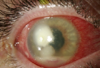
Acanthamoeba keratitis
The protozoan, Acanthamoeba, causes a particularly difficult infection to treat. Acanthamoeba
is found widely in nature and has been isolated from random samples
of soil, water, and air. It can gain access to contact lens solutions
and contact lenses from any of these sources, but tap water is a
common culprit. Tap water should never be used to rinse contact
lenses, store contact lenses, or make saline solution because of the
risk of Acanthamoeba infection.4,5,6,7
The symptoms of infection occur more gradually than in bacterial
keratitis. It often takes several days or weeks before the symptoms
progress to the point that the patient seeks attention. The early signs
and symptoms are foreign body sensation, mild blurred vision, and
redness. This progresses to pain, conjunctival injection, rough
epithelium, and thickened corneal nerves on slit lamp examination. As
the infection progresses, the pain becomes severe, out of proportion to
the apparent amount of inflammation, and a characteristic central
ring infiltrate forms.
Acanthamoeba feed on bacteria and are cultured on a lawn of Escherichia coli. Corneal scrapings or biopsy specimens are placed on the E coli
-layered agar and inspected for the characteristic tracks that the
trophozoites make as they eat their way across the dish. Diagnosis and
therapy are both aided by scraping a large amount of epithelium for
culture. Acanthamoeba cysts also can be identified from corneal scrapings using Giemsa and calcofluor white stains.
The Acanthamoeba organism exists in 2 states, motile
trophozoite and dormant cyst. The cyst form has great resistance to
amebicidal drugs and can lie dormant in the cornea for months.
Therefore, topical antimicrobials are started at a high frequency of
every hour and reduced according to the severity of the toxicity and
symptoms. The traditional treatment of propamidine (Brolene) and
neomycin (Neosporin), supplemented with miconazole, clotrimazole, and
oral ketoconazole, is being replaced by polyhexamethylene biguanide
(PHMB). In a concentration of 0.02%, PHMB is effective in killing both
cysts and trophozoites of many different strains and displays
relatively little corneal toxicity. Therapy is continued every 1-2
hours until clinical improvement is evident, which often is 1-2 weeks.
The frequency of dosing is gradually reduced to 4 times a day.
Treatment usually is continued for a number of months until all
inflammation has resolved.
Therapy with PHMB is complicated by the lack of commercially
available solution. PHMB is not licensed for ocular use in any
country. However, the compound is readily available, and several
pharmacies are willing to formulate the proper dosage and to ship the
preparation.


Three patients with Acanthamoeba keratitis were
successfully treated with a topical application of 0.1% riboflavin
solution and 30 minutes of UV irradiation focused on the corneal
ulcer.8
Prognosis depends greatly on how early in the course of the
disease the infection is diagnosed and therapy is instituted. If
caught early, medical cures are usual. However, if it progresses to
the ring infiltrate stage, which typically takes about 6 weeks, a
medical cure is quite difficult and surgery may be required. Amoebae
other than Acanthamoeba can also cause an infectious keratitis; Vahlkampfia jugosa and Naegleria species have been cultured from the contact lenses of patients with keratitis.
Lastly, in the August 2006 issue of the American Journal of Ophthalmology, Joslin et al reported on an Acanthamoeba
keratitis outbreak in the Chicago area. They postulated that changes
in water quality standards from the Environmental Protection Agency
(EPA) may have played a role in this outbreak. If correct, this may
portend an upsurge in such cases nationally.
Tight lens (acute red eye) syndrome
A contact lens occasionally can become tightly adherent to the eye
and produce marked, diffuse stromal inflammation and an anterior
chamber reaction. The resultant pain, photophobia, injection, and
tearing are typically acute and severe. The epithelium has punctate
staining, diffuse or peripheral infiltrates in the anterior stroma.
The symptoms resolve with removal of the contact lens; the infiltrates
may take a few days to disappear. A short course of topical steroids
will speed the resolution of the symptoms.
Corneal warpage
Prolonged contact lens wear may produce gradual and unpredictable
changes in the contour of the cornea-corneal warpage. The astigmatism
or the general steepness may be either increased or decreased.
Typically, corneal warpage produces an irregular astigmatism, which
reduces the best spectacle correction. Corneal warpage commonly is seen
with hard lenses but also can occur with soft contact lens wear.
Corneas usually regain a stable and regular shape after discontinuation
of the contact lenses, but it may take weeks or even months.
Contact lens–induced keratoconus
The possibility of a causal relationship between keratoconus
and contact lenses is controversial. A high percentage (20-30%) of
patients with keratoconus have worn contact lenses at the time of
diagnosis; however, no causal connection may exist because patients with
keratoconus tend to have myopia and irregular astigmatism and they
would be expected to prefer rigid contact lenses to spectacles.
The strongest evidence to support the hypothesis that wearing
contact lenses can cause keratoconus in certain susceptible
individuals has been presented by Macsai and colleagues.9 In
their series of patients with keratoconus, a difference existed in
those who wore contact lenses at the time of diagnosis (eg, older age,
flatter corneas, more superior cones) than those who wore glasses when
diagnosed. They also reported 2 patients who wore contact lenses in
only 1 eye and developed keratoconus in only that eye. This evidence
is very suggestive that long-term wear of contact lenses can produce
keratoconus in susceptible individuals.
The concept that structural changes in the corneal stroma can be
produced by contact lenses is supported by studies, which have found a
correlation between a reduced thickness, an increased steepness, and a
greater irregularity in the corneas of long-term contact lens wearers
compared to normals. Furthermore, keratocyte density appears to be
reduced in contact lens wearers.
The myopic progression found in adolescents does not appear to be
influenced by contact lens wear. In this study, 175 adolescents aged
11-14 years were assigned randomly to spectacle or contact lens wear.
After 3 years, no difference was noted in spherical equivalent change.
Contact lens wear did not increase or decrease the amount of myopia.
Corneal Endothelium and Summary

Related Subject
ABC of Eyes (ABC Series)
Becker-Shaffer's Diagnosis and Therapy of the Glaucomas
Borish's Clinical Refraction (Benjamin, Borish's Clinical Refraction)
Ophthalmology Atlas Test Yourself Atlas in Ophthalmology
Pearls of Glaucoma Management - Feb 2010 Edition

Share
Back to top Go down
john

john

Membership NO : 1
Male Posts : 1672
Join date : 2011-03-27

Contact Lens Complications Empty
PostSubject: Re: Contact Lens Complications   Contact Lens Complications Icon_minitimeMon Mar 28, 2011 10:53 am

Corneal endothelium
Contact lens wear also may compromise the corneal endothelium.
Wearers have a greater variation in endothelial cell size
(polymegethism) and an increased frequency of nonhexagonal cells
(polymorphism) than do nonwearers. Along with the dramatic alteration
in endothelial cell morphology, a small decrease in endothelial cell
density also has been found in long-term contact lens wearers in soft
and PMMA lenses and in RGP lenses.
Deswelling rates are reduced in contact lens wearers and may
indicate a compromised pump reserve or increased endothelial cell
permeability in these corneas. However, using fluorophotometric
methodology, Bourne et al were unable to demonstrate any significant
differences in permeability or pump function in contact lens wearers.
Therefore, it appears clear that morphological changes occur in the
endothelium with contact lens wear. However, it is not clear that this
difference in morphology is translated to a difference in function.
Summary
All eyes are altered by contact lens wear. Abnormalities of the
lids, conjunctiva, epithelium, stroma, and endothelium are produced.
The changes run the gamut from incidental findings without any
apparent functional significance to severely painful and
sight-threatening pathology. Fortunately, the large majority of
contact lens wearers enjoy the benefits of comfort and excellent
vision without experiencing any significant ill effects. The more we
learn about the complications of contact lens wear, the more we can
help our patients treat and avoid them.
Patient education
For excellent patient education resources, visit eMedicine's Eye and Vision Center. Also, see eMedicine's patient education article Contact Lenses.
When the Solution is the Problem

In the past 5 years, contact lens solutions have been implicated
in health risks for soft contact lens wearers. The incidence of
contact lens–related infections has been on the rise. Any delay in
diagnosis of unexpected fungal and Acanthamoeba infections can
contribute to vision loss. The CDC has been involved in
recommendations for eye care practitioners and patients. While patient
error in proper contact lens care remains a significant factor in eye
infections, the Fusarium keratitis outbreak was also linked to
a contact lens solution (Bausch & Lomb ReNu MoistureLoc. The
following is a review of the Fusarium keratitis outbreak, as well as a discussion of keratitis that results from improper lens disinfection due to patient error.

Poor patient compliance with lens wear schedules and care
regimens has been documented to contribute to eye infections. An
estimated 25% of patients do not always wash their hands prior to
handling contact lenses, and 12% never do. Studies estimate that
40-80% of contact lens wearers do not regularly follow the recommended
lens care regimens.

Even an experienced contact lens wearer is not immune to error in
judgment regarding lens care. For example, a 38-year-old male with
keratoconus reported to clinic with symptoms of blur and discomfort in
the left eye, along with a cloud present with and without the contact
lens. This patient had been a successful SoftPerm lens wearer for the
past 13 years without incident. His health history is significant for
type II diabetes. For nearly 3 months, this patient had difficulty
finding the AOSept disinfection system in local stores. He used only
saline solution and began to notice symptoms of blur after 1 month. He
continued with only saline solution until he finally called the
authors’ office. He was told to use an alternative peroxide system and
come in immediately.

Even after 13 years of contact lens wear, this patient did not
understand the importance of disinfecting his contact lenses. He
realized that there was a cloud that continued when his lens was
removed. Once he knew that something was wrong with his eye itself, he
still waited several weeks to call for an appointment.

The patient’s vision had dropped from his habitual 20/20 to 20/30
in the left eye. Slit lamp examination showed a central stromal scar
and significant corneal neovascularization (see images below).
<blockquote>Contact Lens Complications 1189694-1193217-1196459-1638610tnStromal scar and corneal neovascularization.

</blockquote>[ CLOSE WINDOW ]<blockquote>Contact Lens Complications 1189694-1193217-1196459-1638610</blockquote>Stromal scar and corneal neovascularization

Related Subject
ABC of Eyes (ABC Series)
Becker-Shaffer's Diagnosis and Therapy of the Glaucomas
Borish's Clinical Refraction (Benjamin, Borish's Clinical Refraction)
Ophthalmology Atlas Test Yourself Atlas in Ophthalmology
Pearls of Glaucoma Management - Feb 2010 Edition

Share
Back to top Go down
john

john

Membership NO : 1
Male Posts : 1672
Join date : 2011-03-27

Contact Lens Complications Empty
PostSubject: Re: Contact Lens Complications   Contact Lens Complications Icon_minitimeMon Mar 28, 2011 10:54 am


Contact Lens Complications 1189694-1193217-1196459-1638612tnClose-up view of stromal scar and corneal neovascularization.

[ CLOSE WINDOW ]
Contact Lens Complications 1189694-1193217-1196459-1638612
Close-up view of stromal scar and corneal neovascularization.


He was prescribed a course of topical antibiotics, and his scar
has faded somewhat in the 2 years following this incident. Eye care
practitioners must stress to patients the importance of contact lens
disinfection. That a patient with 13 years of contact lens experience
did not recognize an urgent situation was surprising. The change in
distribution of his usual lens care product, AOSept, led to his using
an inappropriate saline solution rather than a disinfecting solution.

While patient error contributed to an eye infection in the situation above, the fungal Fusarium
outbreak in 2005-2006 was connected to the Bausch & Lomb soft
contact lens solution ReNu with MoistureLoc. In a typical year, an
estimated 21 cases of Fusarium keratitis are reported to the
Centers for Disease Control (CDC). Most of these are in southern
climates, and 2% affect soft contact lens wearers. In 2005, 39 cases of Fusarium
keratitis were reported in soft contact lens wearers. All but 5 of
the 39 had used the ReNu with MoistureLoc solution (originating in a
Bausch & Lomb plant in Greenville, NC).

On April 13, 2006, Bausch & Lomb voluntarily withdrew all
ReNu with MoistureLoc solution and recommended that consumers stop
using the product immediately. Other types of ReNu remained on the
market, as they do today. On May 9, 2006, the CDC had 106 confirmed
cases and 80 still under investigation in a total of 32 states.
Additional cases were reported in Malaysia, Singapore, and Australia.
Treatment varied from topical antifungal agents to intravitreal
injections to corneal transplants, depending on the severity of the
condition and the delay in diagnosis.

CDC recommendations to healthcare providers in May 2006 were as follows:


  • Advise patients to stop using ReNu with MoistureLoc immediately.
  • If a patient’s microbial keratitis persists, consider that fungal
    infection may be involved, especially if the patient wears soft
    contact lenses.
  • Prior to initiating treatment, obtain a specimen for culture.
  • Report cases of fungal keratitis to the FDA.
On August 22, 2006, the CDC announced that users of ReNu with MoistureLoc were 20 times more likely to develop Fusarium keratitis than were contact lens wearers who used other solutions. Between June 1, 2005, and June 30, 2006, 164 cases of Fusarium
keratitis had been confirmed. Of these confirmed cases, 34% of those
people needed a corneal transplant. In 25% of the cases, doctors had
mistakenly prescribed an anti-inflammatory medication that made the Fusarium
keratitisworse. While many patients regained vision, some had
permanent vision loss. Several cases required long courses of treatment
and repeat corneal transplants.

A 2006 article in The New York Times included the
following: "Federal disease control experts and leading eye doctors
have formally concluded that Bausch & Lombs’s ReNu with
MoistureLoc was the only contact lens solution contributing to an
outbreak of potentially blinding fungal eye infections earlier this
year. But the researchers’ report, to be published Wednesday in the
Journal of the American Medical Association, says it remains unclear
how the product caused the problems."10

Legal cases resulting from this outbreak were handled in the
South Carolina court system because the worldwide distribution plant
for ReNu with MoistureLoc is located in that state. Two years later,
1,000 product liability lawsuits had been filed over Renu with
MoistureLoc, possibly costing more than $1 billion in damages.


Contact Lens Complications Progress

Share
Back to top Go down
john

john

Membership NO : 1
Male Posts : 1672
Join date : 2011-03-27

Contact Lens Complications Empty
PostSubject: Re: Contact Lens Complications   Contact Lens Complications Icon_minitimeMon Mar 28, 2011 10:54 am

Just a short while
later, in May of 2007, risks of Acanthamoeba keratitis hit the news.
Advanced Medical Optics (AMO) voluntarily recalled Complete
MoisturePlus multipurpose solution. This time, the concern was Acanthamoeba
keratitis. This is a rare, serious infection caused by a parasite
found in water environments such as pools and lakes (see images
below).<blockquote>Contact Lens Complications 1189694-1193217-1196459-1638615tnAcanthamoeba infection.

</blockquote>[ CLOSE WINDOW ]<blockquote>Contact Lens Complications 1189694-1193217-1196459-1638615</blockquote>Acanthamoeba infection.


<blockquote>Contact Lens Complications 1189694-1193217-1196459-1638616tnAcanthamoeba infection.

</blockquote>[ CLOSE WINDOW ]<blockquote>Contact Lens Complications 1189694-1193217-1196459-1638616</blockquote>Acanthamoeba infection.


<blockquote>Contact Lens Complications 1189694-1193217-1196459-1638618tnCorneal transplant following acanthamoeba infection.

</blockquote>[ CLOSE WINDOW ]<blockquote>Contact Lens Complications 1189694-1193217-1196459-1638618</blockquote>Corneal transplant following acanthamoeba infection.


The CDC had interviewed 46 people who had Acanthamoeba
keratitis between 2005 and 2007. Of these interviewees, 39 were contact
lens wearers, and 21 had reported use of Complete MoisturePlus
multipurpose solution in the month prior to symptoms. No cause and
effect were determined, and products in the marketplace now do not seem
to cause increased risk of infection.

Whether caused by patient error or contact lens solutions, eye
infection rates have caught national attention, and eye care
professionals must take extra care in educating their patients on
proper lens care. Agencies such as the FDA and CDC keep practitioners
and the public informed of any harmful trends. The www.CDC.org
Web site provides guidelines and advice for both consumers and
healthcare providers. Practitioners should ask their patients about
their lens care habits and not assume that patients know the proper
care regimen. Remember that the patient may be not following
instructions, and stay abreast of any product recalls. The solution is
sometimes the problem

Related Subject
ABC of Eyes (ABC Series)
Becker-Shaffer's Diagnosis and Therapy of the Glaucomas
Borish's Clinical Refraction (Benjamin, Borish's Clinical Refraction)
Ophthalmology Atlas Test Yourself Atlas in Ophthalmology
Pearls of Glaucoma Management - Feb 2010 Edition
Contact Lens Complications Progress

Share
Back to top Go down
john

john

Membership NO : 1
Male Posts : 1672
Join date : 2011-03-27

Contact Lens Complications Empty
PostSubject: Re: Contact Lens Complications   Contact Lens Complications Icon_minitimeMon Mar 28, 2011 10:55 am

Media file 1: Stromal scar and corneal neovascularization.[ CLOSE WINDOW ]<blockquote>Contact Lens Complications 1189694-1193217-1196459-1638610</blockquote>Stromal scar and corneal neovascularization.


Contact Lens Complications 1189694-1193217-1196459-1638612tn(Enlarge Image)
Media file 2: Close-up view of stromal scar and corneal neovascularization.
[ CLOSE WINDOW ]<blockquote>Contact Lens Complications 1189694-1193217-1196459-1638612</blockquote>Close-up view of stromal scar and corneal neovascularization.


Contact Lens Complications 1189694-1193217-1196459-1638615tn(Enlarge Image)
Media file 3: Acanthamoeba infection.
[ CLOSE WINDOW ]<blockquote>Contact Lens Complications 1189694-1193217-1196459-1638615</blockquote>Acanthamoeba infection.


Contact Lens Complications 1189694-1193217-1196459-1638616tn(Enlarge Image)
Media file 4: Acanthamoeba infection.
[ CLOSE WINDOW ]<blockquote>Contact Lens Complications 1189694-1193217-1196459-1638616</blockquote>Acanthamoeba infection.


Contact Lens Complications 1189694-1193217-1196459-1638618tn(Enlarge Image)
Media file 5: Cor
Related Subject
ABC of Eyes (ABC Series)
Becker-Shaffer's Diagnosis and Therapy of the Glaucomas
Borish's Clinical Refraction (Benjamin, Borish's Clinical Refraction)
Ophthalmology Atlas Test Yourself Atlas in Ophthalmology
Pearls of Glaucoma Management - Feb 2010 Edition
Contact Lens Complications Progress

Share
Back to top Go down
 

Contact Lens Complications

View previous topic View next topic Back to top 
Page 1 of 1

 Similar topics

-
» Contact Lens Diaphragm
» Contact Dermatitis – November 2010 Edition
» FeverWatch Professional Clinical Non-contact Infrared Thermometer - Forehead
» Complications in Cutaneous Surgery
» Complications of Regional Anesthesia

Permissions in this forum:You cannot reply to topics in this forum
Medical Book :: Clinical Sciences Books :: Ophthalmology-