Malignant Mesothelioma Imaging Mesothelioma,pleural,Mesothelioma,malignant,Mesothelioma
Mesothelioma,malignant,neoplasm,Mesothelioma,pleural,Mesothelioma
peritoneal Mesothelioma,Mesothelioma,neoplasm,Mesothelioma,asbestosis,Mesothelioma
neoplasm,Mesothelioma,peritoneuim,Mesothelioma
OverviewMesothelioma is a malignant neoplasm originating from pleural or peritoneal surfaces; this condition is usually associated with occupational exposure to asbestos. Wagner et al connected asbestos to mesothelioma
in a classic 1960 study of 33 patients with mesothelioma who were
exposed to asbestos in a mining area in South Africa's North Western
Cape Province.
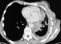
[1] Of the 33 patients, 32 had been exposed to crocidolite, the most carcinogenic type of asbestos.A positron emission tomography (PET) scan of mesothelioma is provided below
.

Positron
emission tomography (PET) scan in a male patient with known
mesothelioma. Although PET scanning is not standard for the evaluation
of mesothelioma, this image illustrates the extent of the disease into
the mediastinum and peritoneum. Asbestos
mining and production peaked from the 1930s-1960s, and asbestos was
used in a variety of products ranging from construction supplies to
brake linings. During World War II, hundreds of thousands of civilian
and military workers, through their occupations, were exposed to asbestos.
Production slowed dramatically in the 1970s as the health risks of
asbestos became known. Governmental restrictions were placed on its use,
and alternative materials became available. Despite these changes,
asbestos continues to be used in the manufacture of some fire safety
products. The clinical latency period between asbestos exposure
and mesothelioma development is 35-40 years, and as a result, the number
of mesothelioma patients has continued to rise despite decreased
asbestos production. The most common findings on physical examination
(79%) are signs of pleural effusion (eg, dullness to percussion, decreased breath sounds). The
diagnosis of mesothelioma should be made with care. A clinical history
of asbestos exposure and radiologic findings that are consistent with
mesothelioma warrant inclusion of mesothelioma in the differential
diagnosis, but it is important to stress that a diagnosis of
mesothelioma cannot be made exclusively with imaging studies. More
common diseases, such as benign asbestos-related pleural disease and
metastatic adenocarcinoma, can look radiographically identical to
mesothelioma. Biopsy with special staining and immunohistochemical and
ultrastructural analysis are absolutely essential for the accurate
diagnosis of mesothelioma. Mesothelioma is very difficult to
treat; the treatment is usually surgical, although other treatment
options such as chemotherapy and radiotherapy are used. The 2 primary
surgical interventions are pleurectomy and extrapleural pneumonectomy
(EPP).
[2]
Preferred examinationChest
radiography is the initial screening examination, while computed
tomography (CT) scanning is preferred for staging the tumor.Magnetic
resonance imaging (MRI) complements CT scanning in some patients. MRI
provides better delineation of soft tissues (better soft-tissue
contrast) and allows imaging in the sagittal and coronal planes.
[3] PET scanning may also be useful in delineating the extent of tumor or metastases.
[4]
Limitations of techniquesChest
radiography has limited usefulness. The radiographic findings of
mesothelioma are nonspecific and are observed in other diseases,
including metastatic carcinoma, lymphoma, and benign asbestos disease.
Small malignant pleural effusions may not be observed on standard
radiographs. Alternatively, large pleural effusions can obscure pleural
thickening or masses; therefore, disease extent is frequently
underestimated in radiographs. CT scanning provides more and
better information than plain radiography with regard to tumor
characteristics and extent. Although MRI is superior to CT scanning in
some areas, this advantage did not change the surgical treatment in a
study by Heelan et al.
[5] Neither
CT scanning nor MRI provides an unequivocal diagnosis of mesothelioma;
tissue biopsy is required for the definitive diagnosis.
Next Section: Radiography
RadiographyThe
most common mesothelioma finding on radiographs is unilateral,
concentric, plaquelike, or nodular pleural thickening (as seen in the
images below). Pleural effusions are common and may obscure the presence
of the underlying pleural thickening. The tumor frequently extends into
the fissures, which become thickened and irregular in contour. A slight
right-sided predominance is observed, possibly because of a larger
pleural surface area. The tumor can rigidly encase the lung, causing
compression of lung parenchyma, diaphragm elevation, intercostal space
narrowing, and mediastinal shift toward the tumor. Calcified pleural
plaques are present in 20% of patients with mesothelioma and are usually
related to the previous asbestos exposure.

Chest
radiograph of a 65-year-old man with left-sided chest pain and
biopsy-proven mesothelioma. The left lateral pleura is thickened and
lobulated, which is often observed with mesothelioma.

Chest
radiograph of a 58-year-old patient with mesothelioma and shortness of
breath. This image reveals diffuse, left-sided pleural thickening, a
pleural effusion, and ipsilateral volume loss. Lung
nodules and hilar masses usually result from direct mesothelioma tumor
extension into the lung parenchyma and mediastinal structures, such as
lymph nodes, the pericardium, and the heart. Mechanical distortion of
the hemithorax, chest wall masses, periosteal rib reaction, or rib
destruction by the tumor are signs of advanced disease. Although usually
unilateral, direct extension of the tumor across the mediastinum into
the contralateral hemithorax does occur.
Degree of confidenceAlthough
a definite diagnosis cannot be made on the basis of plain film
findings, new unilateral pleural thickening or effusion in a patient who
has a history of exposure to asbestos is highly suggestive of
mesothelioma.
Previous
Computed TomographyCT scan findings (examples of which are shown in the images below) are
similar to those of plain films but are seen better and in more detail.
[6, 7] Furthermore,
pleural thickening and effusion can be distinguished with CT scanning.
Nodular pleural thickening, pleural thickening greater than 1 cm,
involvement of the mediastinal pleural surface, and concentric pleural
thickening are all highly suggestive of malignant pleural disease,
either mesothelioma or metastases. The tumor extent along the pleural
surfaces and into the mediastinum, diaphragm, or chest wall can be
evaluated much better with CT scanning than with plain radiography.
Chest wall invasion manifests as obliteration of fat planes or chest
wall nodules. Diaphragmatic invasion, ascites, and omental caking are
common CT scan findings of peritoneal mesothelioma.

Computed
tomography scan of a 58-year-old patient with mesothelioma and
shortness of breath. This image shows the extensive pleural thickening
that is characteristic of mesothelioma, effusion, and reduction in the
volume of the affected hemithorax.
Computed
tomography scan of the chest. This image demonstrates mesothelioma that
extends into the chest wall. Note the concentric left pleural
thickening, pleural effusion, reduction in volume of the left
hemithorax, and the tumor nodules within the chest wall.

Computed
tomography scan in a 48-year-old man with right-sided chest pain and a
"tight sensation," who worked as a welder in a Norfolk, Virginia,
shipyard. This image shows that the thick inhomogeneous pleural rind
encases the lung (causing volume loss) and extends into the major
fissure.

Computed
tomography scan in a 70-year-old man with chronic cough, hoarseness,
and a 20-lb weight loss over 3-4 months. This image demonstrates the
left lung is surrounded by a thickened pleura.

Computed
tomography scan in a 71-year-old man with increasing dyspnea and a
history of asbestos exposure several decades earlier. This image shows
that the right lung is reduced in volume as a result of the encasing
pleural rind. An associated pleural effusion and right lower-lobe
rounded atelectasis are also seen.

Computed
tomography (CT) scan in a male Veterans Administration patient with a
history of asbestos exposure and an enlarging abdominal girth. This
upper CT scan slice reveals the calcified pleural plaques along the
diaphragmatic surface that are associated with asbestos exposure.
Ascites is seen lateral to the liver. Aspiration of the ascitic fluid
demonstrated mesothelioma
.

Computed
tomography (CT) scan in a male Veterans Administration patient. This
lower CT scan slice demonstrates ascites, omental caking, and mesenteric
thickening.
False positives/negativesBenign
pleural plaques or pleural thickening from asbestos exposure may mimic
the appearance of nodular pleural thickening in patients with
mesothelioma (see the image below).

Computed
tomography scan in a 68-year-old man with known asbestos exposure.
Multiple biopsies were negative for mesothelioma, and the chest findings
were attributed to benign, asbestos-related pleural disease, which is a
diagnosis of exclusion.
Previous
Magnetic Resonance ImagingMRI produces images (an example is of which is shown below) in multiple
planes and is superior to CT scanning in demonstrating solitary foci of
chest wall invasion, endothoracic fascial involvement, and diaphragmatic
invasion.
[8, 7] Mesothelioma images on MRI demonstrate minimally increased T1 signals relative to
the chest wall musculature and moderately increased signals on
T2-weighted images or T1-weighted images that have been obtained
following injection of gadolinium. Fibrous pleural plaques are usually
isointense or less intense relative to muscle. Inflammatory pleural
disease may mimic the increased MRI signal intensity of
mesothelioma
.

Magnetic
resonance imaging (MRI) scan in a 72-year-old Veterans Administration
patient with left-sided mesothelioma. Note that the MRI scan well
delineates the soft tissues and, in particular, the thoracoabdominal
interface at the diaphragm. Gadolinium-based
contrast agents (gadopentetate dimeglumine [Magnevist], gadobenate
dimeglumine [MultiHance], gadodiamide [Omniscan], gadoversetamide
[OptiMARK], gadoteridol [ProHance]) have been linked to the development
of nephrogenic systemic fibrosis (NSF) or nephrogenic fibrosing
dermopathy (NFD). The disease has occurred in patients with moderate to
end-stage renal disease after being given a gadolinium-based contrast
agent to enhance MRI or MRA scans.NSF/NFD is a debilitating and
sometimes fatal disease. Characteristics include red or dark patches on
the skin; burning, itching, swelling, hardening, and tightening of the
skin; yellow spots on the whites of the eyes; joint stiffness with
trouble moving or straightening the arms, hands, legs, or feet; pain
deep in the hip bones or ribs; and muscle weakness.
Previous
Next Section: Radiography
UltrasonographyUltrasonography
can demonstrate pleural thickening or effusions in patients with
mesothelioma. This modality can be used as a guide for biopsy, but it is
not typically used to assess the extent of disease in patients with
mesothelioma.
Previous
Next Section: Radiography
Nuclear ImagingIf
surgical resection of the tumor is a possibility, a quantitative
ventilation-perfusion scan helps in assessing the function of the
contralateral lung. PET scanning has been used, although not
routinely, to evaluate mesothelioma and may help preoperatively by
documenting the extent of lymph node involvement or distant metastases.
[3, 4, 9, 10, 11, 12] Yildirim
et al examined the efficacy of using
2-[fluorine-18]fluoro-2-deoxy-D-glucose (FDG) PET and CT scanning
together to differentiate malignant mesothelioma from asbestos-related
benign pleural disease. In a study of 31 patients (17 with malignant
mesotheliomas, 9 with benign asbestos pleurisies, 5 with diffuse pleural
fibrosis), the authors found that FDG PET/CT scanning accurately
detected malignant lesions in 15 of the 17 patients with these neoplasms
and that the combined modalities had a sensitivity of 88.2%,
specificity of 92.9%, and overall accuracy of 90.3%. In addition, benign
pleural disease was correctly detected in 13 of 14 patients.
[9] A
study by Mavi et al concluded that dual time-point FDG PET scanning
seems to be an accurate means of differentiating malignant mesothelioma
from benign pleural disease. In the study, which involved 55 patients,
the authors found that at the second time point, FDG uptake in malignant
lesions had increased over that at the first time point, while in
benign lesions, uptake at the second time point had generally remained
stable or had decreased.
[10] Evidence
from a similar study by Yamamoto et al also suggested that dual
time-point FDG is useful for differentiating malignant pleural
mesothelioma from benign pleural lesions.
[11]
False positives/negativesPleural inflammation can also reveal increased uptake on PET scanning.


